Stages of NK and clinical presentation
KNOW THE CONSEQUENCES OF NK
Neurotrophic keratitis (NK) may progress from mild to moderate to severe, and may ultimately lead to corneal perforation, stromal melting, and vision loss.1,2
The image is for illustrative purposes only.
The image is for illustrative purposes only.
NK Damage
NK DAMAGE
SEE THE DAMAGE OF NK
Take a detailed look at the corneal damage that may occur at each stage of neurotrophic keratitis (NK).
A full transcript of the video is available here.
ROOT PATHOGENESIS
ROOT PATHOGENESIS
A DEGENERATIVE DISEASE THAT MAY BE CAUSED BY CORNEAL NERVE DAMAGE
Neurotrophic keratitis (NK) is defined as a rare disease, and can be mistaken for more common conditions, such as dry eye disease.1,3
A key differentiator between NK and other ocular conditions is the reduction in or total loss of corneal sensitivity.1 This loss may be caused by damage to the corneal nerves, which may lead to poor healing and deterioration of the corneal epithelium.1,4
Other characteristics of disease may include decreased tear production and tear film instability, and decreased blink rate.1,4 Keep reading to learn more about characterization of each stage of disease.
The image is for illustrative purposes only.
DISEASE CASCADE
DISEASE CASCADE
IMPAIRED CORNEAL NERVE FUNCTION SETS OFF A CASCADE OF EVENTS
Multiple etiologies can lead to impairment of the corneal nerves.1 Damage to the corneal nerves reduces the eyes’ ability to maintain ocular surface integrity.4 The loss of corneal innervation leads to a cascade of events resulting in spontaneous corneal epithelial breakdown, which can lead to neurotrophic keratitis (NK).2,4
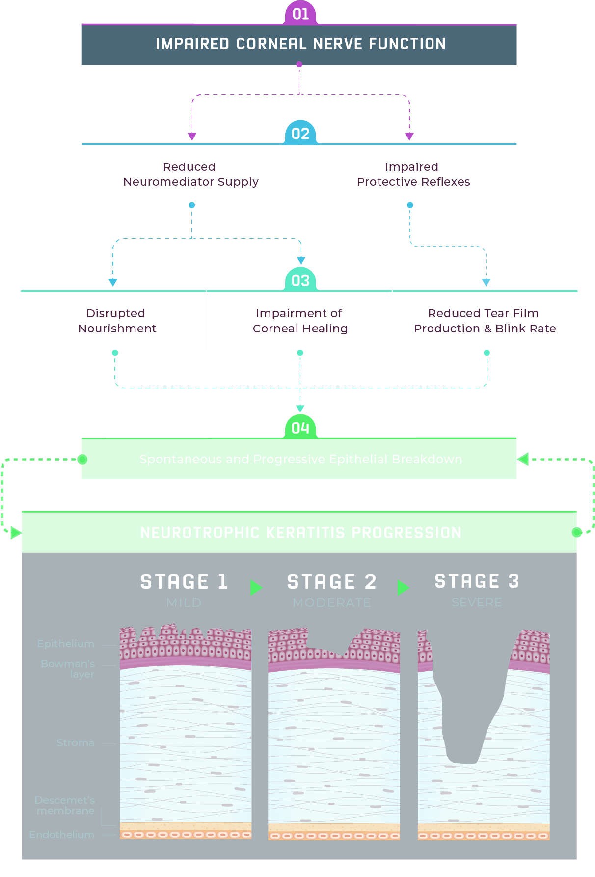

Impairment of corneal nerves prompts a series of events that may cause progressive damage.4

A reduction in neuromediator supply may lead to impairment of neurotrophic factors and a decreased corneal healing rate.4
Corneal nerve impairment may also cause impaired protective reflexes (reduced tear film production and blink rate).4

The combination of disrupted nourishment and reduced corneal healing, tear film production, and blink rate may contribute to the spontaneous breakdown of the corneal epithelium.4

Spontaneous corneal epithelial breakdown may lead to NK, ranging from an irregular, dry, and cloudy corneal epithelium or punctate keratitis (Stage 1), to persistent epithelial defects (Stage 2), and corneal ulcers, stromal melting, or perforation (Stage 3).2,4
Stages of NK
Stages of NK
Assessing severity with Mackie and Dua Classifications
Neurotrophic keratitis (NK) staging is based on the severity of corneal epithelial and stromal involvement, ranging from superficial punctate keratopathy to persistent epithelial defects and ulcers.1 Mackie classification is used to categorize NK in 3 stages.1 Dua classification describes these stages as mild, moderate, and severe.1
Fluorescein staining can help identify severity of disease—if staining reveals significant epithelial damage and the patient does not report any ocular discomfort, it may be a sign of NK.1,3
Mild to moderate NK may progress to severe disease with the potential for vision loss resulting from scarring and corneal perforation.1
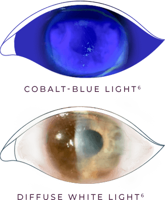
STAGE 1 (MILD)
STAGE 1 (MILD) is characterized by corneal epithelial changes with dry and cloudy corneal epithelium, and the presence of superficial punctate keratitis (SPK).1,2
These images are for illustrative purposes only. These are not an exact representation of a patient, or a structure and function.
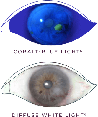
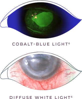
STAGE 2 (MODERATE)
STAGE 2 (MODERATE) is characterized by recurrent and/or persistent epithelial defects (PED) with an oval or circular shape, most frequently located paracentrally on the cornea.1
These images are for illustrative purposes only. These are not an exact representation of a patient, or a structure and function.
STAGE 3 (SEVERE)
STAGE 3 (SEVERE) is characterized by corneal ulcer with stromal involvement or melting that may progress to corneal perforation.1
These images are for illustrative purposes only. These are not an exact representation of a patient, or a structure and function.
STAGE 1 (MILD)
STAGE 1 (MILD) is characterized by corneal epithelial changes with dry and cloudy corneal epithelium, and the presence of superficial punctate keratitis (SPK).1,2
These images are for illustrative purposes only. These are not an exact representation of a patient, or a structure and function.
STAGE 2 (MODERATE)
STAGE 2 (MODERATE) is characterized by recurrent and/or persistent epithelial defects (PED) with an oval or circular shape, most frequently located paracentrally on the cornea.1
These images are for illustrative purposes only. These are not an exact representation of a patient, or a structure and function.
STAGE 3 (SEVERE)
STAGE 3 (SEVERE) is characterized by corneal ulcer with stromal involvement or melting that may progress to corneal perforation.1
These images are for illustrative purposes only. These are not an exact representation of a patient, or a structure and function.
References: 1. Dua HS, Said DG, Messmer EM, et al. Neurotrophic keratopathy. Prog Retin Eye Res. 2018;66:107-131. 2. Sacchetti M, Lambiase A. Diagnosis and management of neurotrophic keratitis. Clin Ophthalmol. 2014;8:571-579. 3. Dana R, Bradley JL, Guerin A, et al. Estimated prevalence and incidence of dry eye disease based on coding analysis of a large, all-age United States health care system. Am J Ophthalmol. 2019;202:47-54. 4. Mastropasqua L, Massaro-Giordano G, Nubile M, Sacchetti M. Understanding the pathogenesis of neurotrophic keratitis: the role of corneal nerves. J Cell Physiol. 2017;232(4):717-724.