Stain without pain?
It may be a sign of neurotrophic keratitis (NK)1
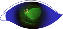
If corneal fluorescein staining reveals epithelial dryness, defects, or ulcers—but the patient does not complain of discomfort due to reduced corneal sensitivity—it may be NK.2
Learn more about this degenerative corneal disease and considerations for making an accurate diagnosis.2
The image is for illustrative purposes only.
RECOGNIZING NK
It may be a sign of neurotrophic keratitis (NK)1
If corneal fluorescein staining reveals epithelial dryness, defects, or ulcers—but the patient does not complain of discomfort due to reduced corneal sensitivity—it may be NK.2
Learn more about this degenerative corneal disease and considerations for making an accurate diagnosis.2
The image is for illustrative purposes only.
RECOGNIZING NK
recognizing nk
NK may be mistaken for other ocular conditions
Neurotrophic keratitis (NK) is a degenerative ocular disease caused by impairment of the corneal nerves and is characterized by a reduction in or loss of corneal sensitivity.2
Early signs of NK may overlap with features of dry eye, and, as a result, it can go unrecognized.2
Evaluating a patient’s medical history and assessing common risk factors are crucial to identifying NK.3 For example, people who have had herpetic eye disease, ocular surgery, or diabetes can be at greater risk of developing NK.3
NK PRESENTATION

Not actual patients.
NK PRESENTATION
THE STAGES OF NK
One way to assess the severity of neurotrophic keratitis (NK) is to use a 3-stage system known as the Mackie classification, later updated by Dua, where severity is characterized by the degree of corneal epithelial and stromal involvement ranging from superficial punctate keratopathy to persistent epithelial defects and ulcers.2,4
Mild (Stage 1) to moderate (Stage 2) NK may progress to severe disease (Stage 3), with risk of vision loss from scarring, corneal melting, and perforation.2,4
These descriptions are not representative of all patients, and individual presentation of NK may vary.

Mild
Stage 1

Moderate
Stage 2
Severe
Stage 3
These images are for illustrative purposes only. These are not an exact representation of a patient, or a structure and function.
Testing for NK
Testing for NK
IF THIS FEELS OK, IT MAY BE NK
For some ocular conditions, eye discomfort or pain is a clear symptom. But in patients with neurotrophic keratitis (NK), decreased corneal sensitivity is a key characteristic.2 “STAIN WITHOUT PAIN” can be an indicator of NK that might otherwise be missed due to reduced or absent corneal sensation.1,2 Corneal sensitivity testing, which can be done with a cotton wisp, can help you determine if a patient has NK.2
The image is for illustrative purposes only.
References: 1. Al-Aqaba MA, Dhillon VK, Mohammed I, et al. Corneal nerves in health and disease. Prog Retin Eye Res. 2019;73:100762. 2. Dua HS, Said DG, Messmer EM, et al. Neurotrophic keratopathy. Prog Retin Eye Res. 2018;66:107-131. 3. Mastropasqua L, Massaro-Giordano G, Nubile M, Sacchetti M. Understanding the pathogenesis of neurotrophic keratitis: the role of corneal nerves. J Cell Physiol. 2017;232(4):717-724. 4. Sacchetti M, Lambiase A. Diagnosis and management of neurotrophic keratitis. Clin Ophthalmol. 2014;8:571-579.